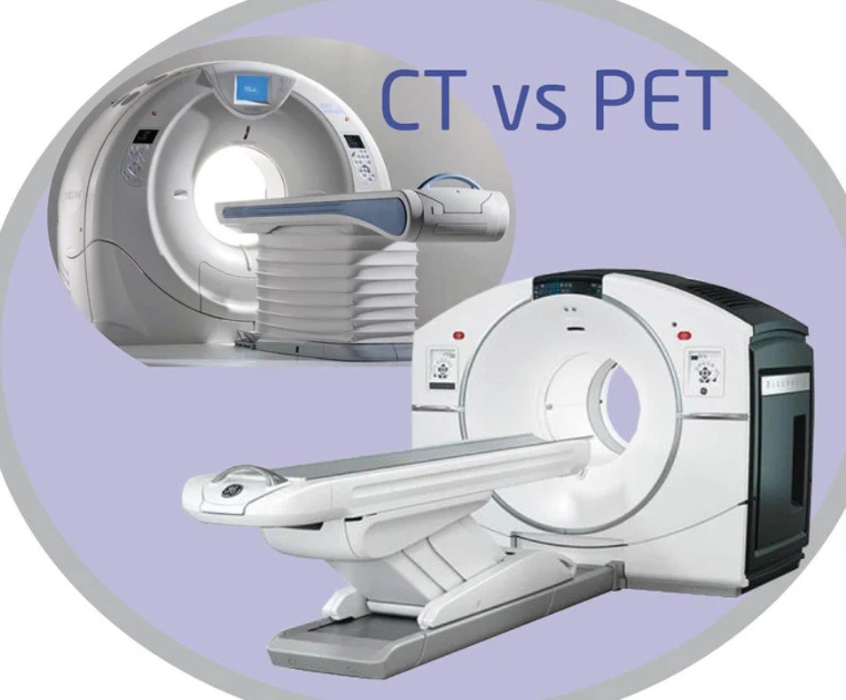
Are you facing a medical condition that requires diagnostic imaging? If so, you may need to undergo a scan using nuclear medicine. This type of scan uses a specialized scan machine to capture images of your body. It is important to note that this procedure involves radiation therapy. Do you find yourself confused about which imaging technique to choose for scanning images? Whether you need to scan images for a scan machine or for radiation therapy, it’s important to consider the best option.

Both PET scans and CT scans play crucial roles. PET scans, also known as positron emission tomography scans, utilize radiotracers to detect metabolic activity in your body. These imaging tests are commonly performed at specialized imaging centers. In contrast, CT scans, or computed tomography scans, use X-rays to create detailed images of internal structures for various medical uses. These imaging tests are commonly employed in medical uses such as cancer detection and staging, as well as evaluating lung conditions and identifying bone fractures. They are often conducted in imaging centers.
By understanding the distinctions between these two imaging methods, you can make an informed decision about the most appropriate option for your needs. Whether you are seeking a provider for cancer treatment or need a doctor to administer a radioactive substance, it is important to have all the necessary information. So let’s dive in and uncover the nuances of PET scans and CT scans, which use radioactive substances to detect cancer. These scans are conducted by doctors and their staff.
Understanding the Differences: Uses, Risks, and Procedure
PET scans and CT scans are both valuable medical imaging techniques that provide doctors with essential information about various medical conditions, including cancer. The medical staff often rely on these scans to accurately diagnose and monitor the progression of the disease. In addition to their diagnostic capabilities, these scans also help identify the need for further treatment options, such as surgery or chemotherapy. It is important for patients to disclose any substance use, as certain substances can interfere with the accuracy of these scans. While imaging scans and cancer have some similarities, there are distinct differences in their uses, procedures, associated risks, and the staff involved.
PET Scan Uses
PET scans are primarily used by medical staff to identify cancerous cells, assess brain disorders, and evaluate heart conditions. The staff can detect changes in cancer cellular activity and metabolism within the body. By injecting a radioactive tracer into the patient’s bloodstream or inhaling it as a gas, doctors can visualize areas of abnormality or disease with the help of staff. PET scans are essential for cancer diagnosis and treatment planning, as they help locate tumors and assess if they have metastasized to other areas of the body. These scans are crucial for medical staff.
CT Scan Uses
On the other hand, CT scans are often employed to diagnose injuries or diseases affecting bones, blood vessels, lungs, abdomen, or pelvis. This imaging technique provides detailed cross-sectional images of these areas by using X-rays from different angles. CT scans help doctors identify fractures, tumors, blood clots, infections, or other abnormalities within the body. They are particularly useful for guiding surgical procedures and monitoring treatment effectiveness.
Procedure for PET Scans
The procedure for a PET scan involves injecting a radioactive tracer into the patient’s bloodstream or inhaling it as a gas. The tracer emits positrons that collide with electrons inside the body tissues. These collisions produce gamma rays that are detected by specialized cameras surrounding the patient during scanning. The data collected is then processed by computers to create detailed images showing areas of high metabolic activity.
During a PET scan procedure:
-
The patient will be asked to lie down on an examination table.
-
A small needle is used to inject the radioactive tracer into a vein.
-
Alternatively, patients may be instructed to inhale a gaseous form of the tracer.
-
After an appropriate waiting period for the tracer to distribute throughout the body, scanning begins.
-
The table moves slowly through the doughnut-shaped PET scanner, capturing images from different angles.
-
The process usually takes about 30-60 minutes to complete.
Procedure for CT Scans
In contrast, a CT scan requires the patient to lie on a table that moves through a doughnut-shaped machine emitting X-rays. The X-ray beams pass through the body and are detected by sensors on the opposite side of the machine. These sensors transmit data to a computer that reconstructs detailed cross-sectional images of the scanned area.
During a CT scan procedure:
-
The patient will be positioned on the examination table.
-
Straps or pillows may be used to help maintain stillness during scanning.
-
The table slowly moves through the doughnut-shaped CT scanner while multiple X-ray beams rotate around it.
-
The process is quick and typically lasts only a few minutes.
Risks and Safety Considerations
Both PET scans and CT scans involve exposure to radiation, but the risks associated with this exposure are generally minimal. Radiologists and technologists take precautions to ensure patients receive only necessary radiation doses while maximizing diagnostic accuracy.
Some individuals may experience allergic reactions to contrast agents used in both types of scans. However, these reactions are rare and can be managed effectively with medications if they occur.
Explaining PET Scans: Types, Purpose, and Results
PET scans, also known as positron emission tomography scans, are a valuable diagnostic tool used in the field of medicine. These scans provide detailed information about the metabolic activity within tissues and help doctors detect abnormalities at the cellular level.
Different Types of PET Scans
PET scans come in various forms depending on their specific purpose. One common type is FDG-PET, which stands for fluorodeoxyglucose positron emission tomography. This scan detects glucose metabolism in tissues and is often used to identify cancerous cells or areas with increased metabolic activity.
Another type of PET scan is Amyloid-PET, primarily employed in diagnosing Alzheimer’s disease. It helps identify amyloid plaques that accumulate in the brains of individuals with this neurodegenerative disorder. By detecting these plaques early on, doctors can initiate appropriate treatment plans.
MIBG-PET is yet another variant used to evaluate neuroendocrine tumors. This scan measures the uptake of metaiodobenzylguanidine (MIBG) by tumor cells and aids in determining the extent and location of such tumors.
Purpose of PET Scans
The main purpose of a PET scan is to detect abnormalities at the cellular level by measuring metabolic activity within tissues. Unlike other imaging techniques like CT scans or X-rays that focus on structural changes, PET scans provide insight into functional changes occurring within organs or systems.
By pinpointing areas with abnormal metabolic activity, doctors can identify potential cancerous cells or other conditions requiring further investigation. For example, if a patient experiences bone pain without an apparent cause visible on an X-ray, a bone-specific PET scan can reveal any underlying abnormalities.
Interpreting PET Scan Results
After undergoing a PET scan procedure, patients receive results that show areas of increased or decreased metabolic activity. These results are often presented in the form of colored images or numerical values. The colors indicate the intensity of metabolic activity, with warmer colors like red indicating higher activity and cooler colors like blue indicating lower activity.
When analyzing these results, doctors look for any significant deviations from normal metabolic patterns. For instance, if a particular region displays heightened metabolic activity, it may suggest the presence of cancerous cells or an inflammatory process.
It is important to note that PET scans alone cannot provide a definitive diagnosis. Instead, they serve as a valuable tool to guide further investigations and help doctors make informed decisions about treatment options.
Exploring CT Scans: Types, Purpose, and Results
CT scans, also known as computed tomography scans, are a valuable medical imaging tool that can provide detailed cross-sectional images of various parts of the body. These scans can be performed with or without contrast agents, depending on the specific area being examined.
The primary purpose of a CT scan is to create high-resolution images that allow doctors to identify and diagnose various conditions affecting bones, blood vessels, organs, or soft tissues. By visualizing these structures in detail, healthcare professionals can gain crucial insights into potential abnormalities or diseases.
After undergoing a CT scan procedure, patients receive their test results in the form of these detailed images. These results play a vital role in helping doctors make accurate diagnoses and develop appropriate treatment plans. The clarity and precision offered by CT scans enable medical professionals to detect fractures, tumors, infections, blood clots, brain disorders, and other abnormalities within the body.
One advantage of CT scans is their versatility. They can be used for different purposes depending on the patient’s needs. For example:
-
In emergency situations where time is critical, a CT scan can quickly provide essential information about injuries sustained from accidents or trauma.
-
When investigating possible brain disorders such as strokes or tumors, a specialized CT scanner tailored for brain imaging may be used.
-
For evaluating chest conditions like lung cancer or pneumonia, a chest CT scan offers detailed views of the lungs and surrounding structures.
-
Abdominal CT scans help identify issues with organs such as the liver, kidneys, pancreas, or intestines.
In terms of convenience for patients undergoing a CT exam using contrast agents (contrast-enhanced), it usually involves an injection before the scan begins. This dye helps highlight certain areas within the body to improve visibility during image interpretation.
CT scanners themselves consist of an examination table that moves through a large circular opening while taking X-ray images from multiple angles. These images are then processed by a computer to create the final cross-sectional images.
Comparing PET Scan and CT Scan: Similarities and Differences
Both PET scans and CT scans are non-invasive imaging techniques used in medical diagnostics. While they serve a similar purpose, there are distinct differences in their methodology and the information they provide.
PET scans, or Positron Emission Tomography scans, offer valuable insights into cellular metabolism and function. They involve the use of radioactive tracers that are injected into the body. These tracers emit positrons, which collide with electrons in the body’s cells, resulting in gamma rays that can be detected by the PET scanner. By analyzing these gamma rays, doctors can assess metabolic activity within various organs and tissues.
On the other hand, CT scans (Computed Tomography scans) provide detailed anatomical images of different parts of the body. This imaging technique utilizes X-rays to create cross-sectional images or slices of the body. By combining multiple slices, doctors can obtain a comprehensive view of specific areas such as the brain, bones, or organs.
The key distinction between PET scans and CT scans lies in their underlying principles. PET scans focus on cellular activity while CT scans concentrate on anatomical structures. This means that PET scans excel at identifying functional abnormalities such as tumors or abnormal cell growths within organs. In contrast, CT scans are particularly effective at visualizing bone structure and detecting fractures or other bone-related issues.
To enhance diagnostic capabilities further, medical professionals often employ a combination of both techniques through a procedure known as PET/CT scanning. By merging functional information from PET with anatomical data from CT, this hybrid approach provides a more comprehensive understanding of a patient’s condition.
During a PET/CT scan procedure, patients receive an injection of radioactive tracer before undergoing both types of imaging sequentially within one machine. The integration allows for precise alignment between functional abnormalities detected by PET and their corresponding anatomical location identified by CT.
In addition to using radioactive tracers for metabolic assessment, PET scans may also involve the administration of contrast liquid to enhance the visibility of specific structures. This is particularly useful when examining blood vessels or certain organs like the heart.
Benefits and Limitations of PET Scan vs CT Scan
PET scans and CT scans are two commonly used diagnostic tools. Both have their own set of benefits and limitations that make them suitable for different situations. Let’s explore the advantages and drawbacks of each.
Benefits of PET Scans
One major benefit of PET scans is their ability to detect diseases at an early stage, even before they become visible on other imaging tests. This is particularly valuable in the case of cancer, where early detection can significantly improve treatment outcomes. By measuring metabolic activity in the body, PET scans can identify areas with abnormal cell growth or high glucose uptake, indicating the presence of tumors or other abnormalities.
Moreover, PET scans provide a comprehensive view of the entire body, allowing doctors to evaluate multiple organs and systems simultaneously. This makes them especially useful for staging cancers and monitoring treatment effectiveness over time. PET scans can help differentiate between benign and malignant tumors, aiding in accurate diagnosis.
However, it’s worth noting that there may be limitations to accessing PET scans due to cost or availability issues. These scans require specialized equipment and radiopharmaceuticals, which can make them more expensive compared to other imaging modalities. Furthermore, not all healthcare facilities have PET scan capabilities, particularly in rural or remote areas.
Benefits of CT Scans
On the other hand, CT scans offer their own unique benefits in medical imaging. One significant advantage is their ability to provide highly detailed images quickly. CT scanners use X-rays from multiple angles to create cross-sectional images of the body’s structures with exceptional clarity. This allows healthcare professionals to visualize intricate details such as blood vessels or small lesions accurately.
Another advantage is that CT scans are widely available in most hospitals and clinics worldwide. Their accessibility makes them a go-to option for various medical conditions requiring detailed anatomical information without delaying diagnosis or treatment plans.
Nevertheless, one limitation associated with CT scans is the exposure to ionizing radiation. Although the radiation doses from modern CT scanners are considered safe, repeated examinations may pose potential risks, especially for younger patients or those who require frequent imaging studies.
Limitations of PET Scans
PET scanning does have its limitations as well. One such limitation is the possibility of false-positive results. Increased metabolic activity caused by inflammation can mimic cancerous cells on a PET scan, leading to unnecessary anxiety and further invasive procedures. This highlights the importance of correlating PET scan findings with other diagnostic tests to confirm accurate diagnoses.
Limitations of CT Scans
Similarly, CT scans also have their own set of limitations. Apart from the potential risks associated with radiation exposure during repeated examinations, some patients may experience claustrophobia or discomfort due to the need for complete stillness during the procedure. Certain individuals with allergies or kidney problems may face challenges when contrast agents are used in conjunction with CT scans.
Addressing Risks and Safety Concerns in PET-CT Imaging
Many patients have concerns about the potential risks involved. While both types of imaging are generally safe, it is important to address any safety concerns and take necessary precautions to ensure patient well-being.
Risks Associated with PET and CT Imaging Procedures
Both PET (Positron Emission Tomography) and CT (Computed Tomography) scans involve the use of radiation. This raises concerns about potential radiation exposure for patients. However, it is essential to note that the radiation dose from these procedures is relatively low and considered safe for diagnostic purposes.
Another risk associated with these imaging procedures is the possibility of allergic reactions to contrast agents. Contrast agents are sometimes used during PET or CT scans to enhance visibility of certain body structures or abnormalities. Although rare, some individuals may be allergic to these agents, resulting in mild to severe reactions. It is crucial for patients to inform their healthcare providers about any known allergies before undergoing a PET-CT scan.
Minimizing Radiation Exposure: Modern CT Scanners’ Role
To minimize radiation exposure during CT scans, modern scanners are equipped with advanced technologies and dose reduction techniques. These advancements allow radiologists and technologists to obtain high-quality images while minimizing the amount of radiation delivered to the patient.
Strict protocols are followed when performing CT scans to ensure that only necessary areas are scanned, reducing unnecessary exposure. The use of low-dose protocols further helps in limiting radiation doses without compromising diagnostic accuracy.
Ensuring Patient Safety: Guidelines Followed by Radiologists and Technologists
Radiologists and technologists who perform PET-CT imaging procedures follow strict safety guidelines to prioritize patient well-being. These guidelines include:
-
Properly assessing each patient’s medical history before proceeding with the scan.
-
Informing patients about the procedure, its benefits, and any potential risks involved.
-
Ensuring that the patient is positioned correctly to obtain accurate images while minimizing discomfort.
-
Monitoring patients throughout the procedure to address any immediate concerns or reactions promptly.
Radiologists and technologists receive specialized training to operate the imaging equipment safely and effectively. This helps in reducing the likelihood of errors and ensuring optimal image quality while maintaining patient safety.
Patient Responsibility: Communicating Allergies and Medical Conditions
Patients also play a crucial role in ensuring their own safety during PET-CT scans. It is essential for individuals to communicate any known allergies or medical conditions to their healthcare providers before undergoing the procedure. By providing this information, medical professionals can take necessary precautions or make appropriate adjustments to ensure patient safety.
Choosing the Right Imaging Technique for Your Needs
Now that you have a better understanding of PET scans and CT scans, you can make an informed decision about which imaging technique is right for your needs. If you’re looking for detailed images of your body’s internal structures, a CT scan may be the best option. On the other hand, if you need to assess the functionality of organs and tissues, a PET scan can provide valuable information. It’s important to consult with your healthcare provider to determine which technique will provide the most accurate diagnosis or evaluation for your specific situation. Don’t hesitate to ask questions and voice any concerns you may have.
FAQs
[faq-schema id=”481″]


